
Introduction to Medical Image Analysis in the 2000s
The 2000s were a transformative time for medical image analysis, marked by a shift from traditional methods to advanced computational techniques. This era was characterized by increased computational power and innovative algorithms that improved diagnostic accuracy and expanded medical applications, leading to more individualized treatment approaches.
Key Technological Developments
Several pivotal advancements defined the technological progress in medical image analysis during this decade:
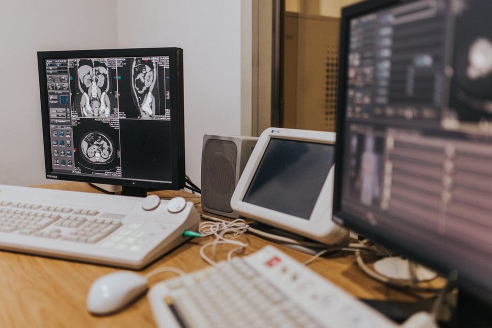
- Advanced Imaging Modalities: Enhancements in MRI, CT, and ultrasound technologies enabled the creation of high-quality images essential for precise analysis. These modalities offered detailed anatomical insights, further refined by improved image processing methods.
- Machine Learning Algorithms: Early machine learning techniques, such as support vector machines and principal component analysis, introduced new possibilities for image classification, segmentation, and pattern recognition. These foundational algorithms paved the way for more sophisticated models in subsequent years.
- Integration of Multi-Modal Data: The ability to combine data from different imaging modalities allowed for a more thorough analysis. This approach improved disease diagnosis and monitoring by correlating diverse types of information.
Applications in Clinical Practice
In the 2000s, medical image analysis had a direct impact on clinical practice. Enhanced algorithms and techniques improved tumor detection and classification, which are critical for cancer diagnosis and treatment planning. Automated systems developed for identifying anomalies in mammograms and other scans reduced radiologists' workload and increased diagnostic precision.
Advancements in image registration techniques also enabled better tracking of disease progression by aligning images taken at different times, a feature particularly useful in monitoring chronic conditions like multiple sclerosis and evaluating treatment effectiveness.
Challenges and Limitations
Despite these advancements, the field faced significant challenges. One major issue was the requirement for large annotated datasets to effectively train machine learning models. The absence of standardized datasets and varying imaging protocols across institutions made it difficult to develop universally applicable solutions.
Additionally, the computational cost of processing large volumes of high-resolution images was a hurdle. Although computational power was on the rise, it remained a limiting factor for the widespread adoption of complex algorithms in routine clinical practice.
Legacy and Impact on Future Developments
The technological strides of the 2000s laid the foundation for future advancements in medical image analysis. The progress in algorithms and computational techniques during this period prepared the ground for the introduction of deep learning in the next decade, which offered even more powerful tools for image analysis.
This decade also highlighted the importance of interdisciplinary collaboration, bringing together experts from computer science, radiology, and clinical medicine to create solutions that were both technically advanced and clinically relevant. The insights gained during this period continue to guide ongoing research and development in medical imaging.
Conclusion
The 2000s were a crucial decade for medical image analysis, with the challenges faced and solutions developed during this time leaving a lasting impact on the field. The technological advancements not only enhanced diagnostic capabilities but also set the stage for future innovations, underscoring the necessity of continued research and development in medical imaging technologies.